3 Things to Know About the Signal Path of the Auditory System — Pro Audio Files
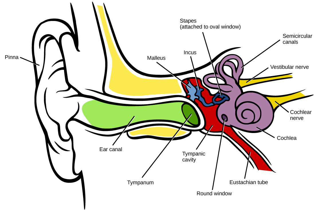
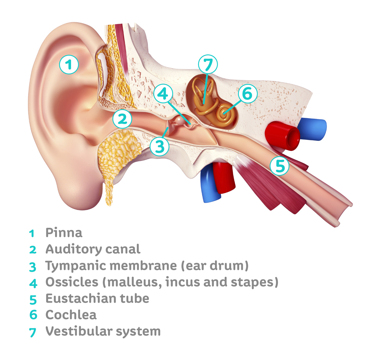
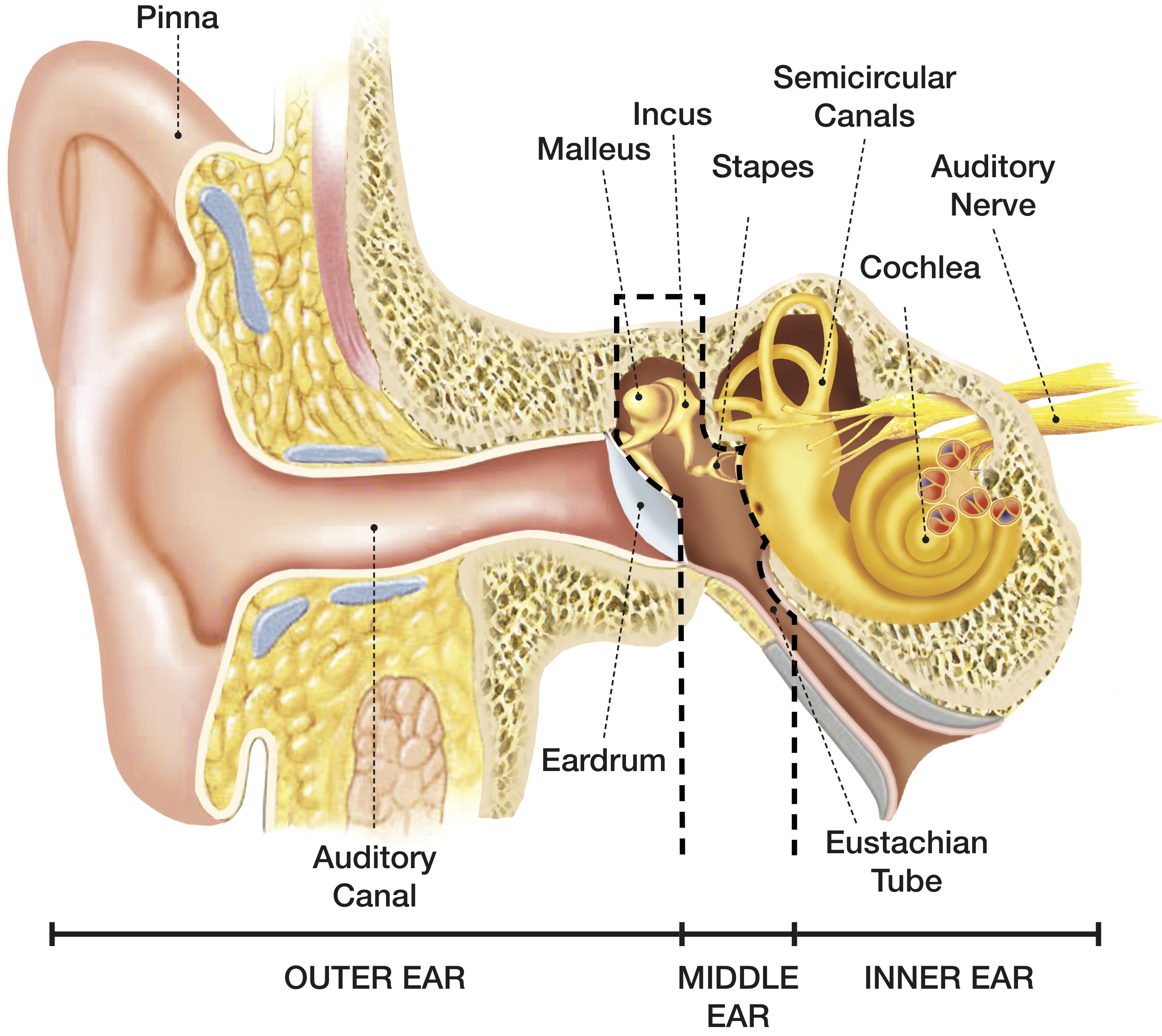
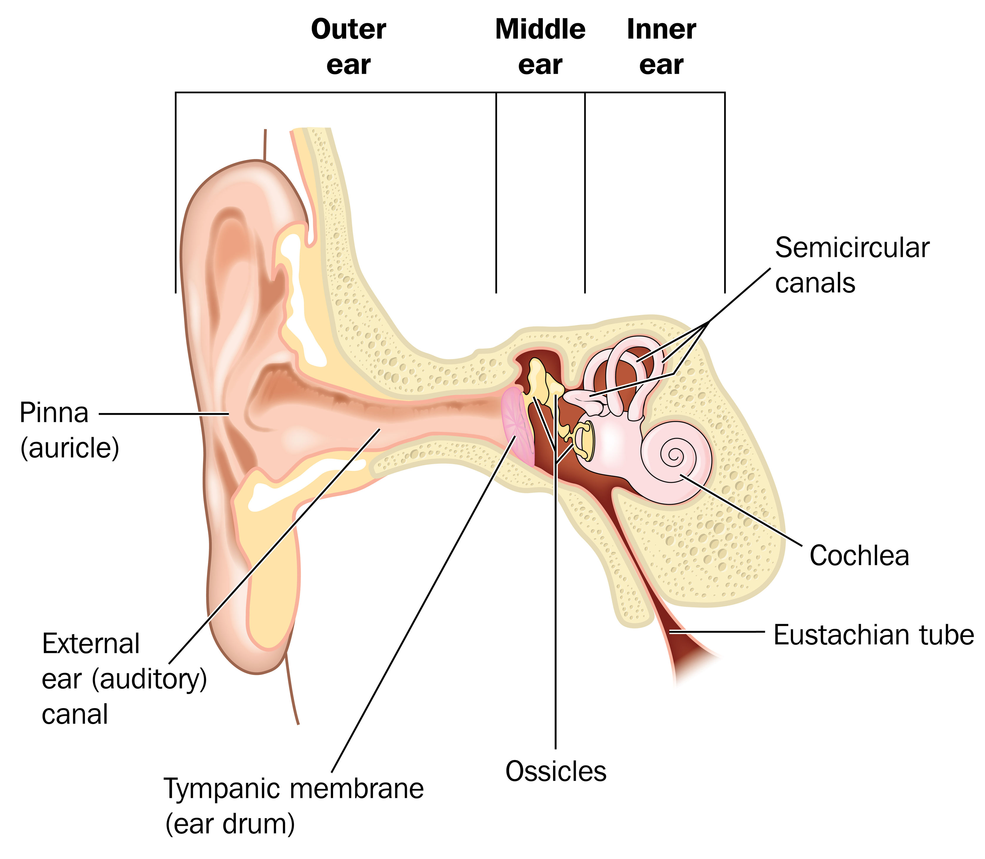
How Do We Hear? The sound waves travel first through the ear canal and vibrate the eardrum. Before the sound waves enter the inner ear, the total pressure must be amplified. The ossicles in the middle ear do the job of amplification. Then the cochlea in the inner ear conducts the sound through a fluid.
The human ear structure and how it works Connect Hearing
human ear, organ of hearing and equilibrium that detects and analyzes sound by transduction (or the conversion of sound waves into electrochemical impulses) and maintains the sense of balance (equilibrium). Understand the science of hearing and how humans and other mammals perceive sound How humans and other mammals perceive sound.
The Ear — Summerlin Audiology
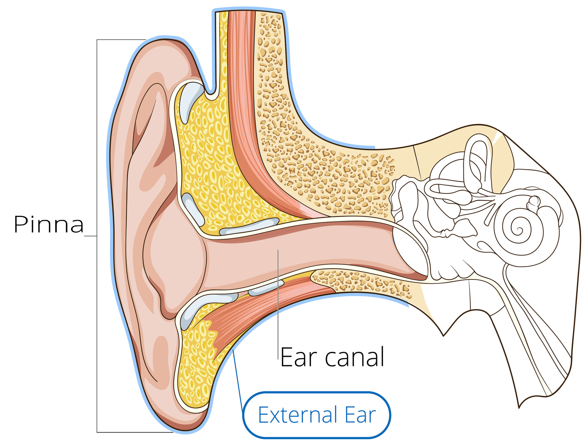
External acoustic meatus. The ear canal, also called the external acoustic meatus, is a passage comprised of bone and skin leading to the eardrum. The ear is comprised of the ear canal (also known.
Hearing Sense Ask A Biologist
Let's explore how human ears work. More free lessons & practice on this chapter-https://www.khanacademy.org/science/in-in-class9th-physics-india/in-in-sound-.

The Anatomy of the Outer Ear Health Life Media
Ear Anatomy, Diagram & Pictures | Body Maps Human body Head Ear Ear The ears are organs that provide two main functions — hearing and balance — that depend on specialized receptors called.
How We Hear Hearing Associates, Inc.
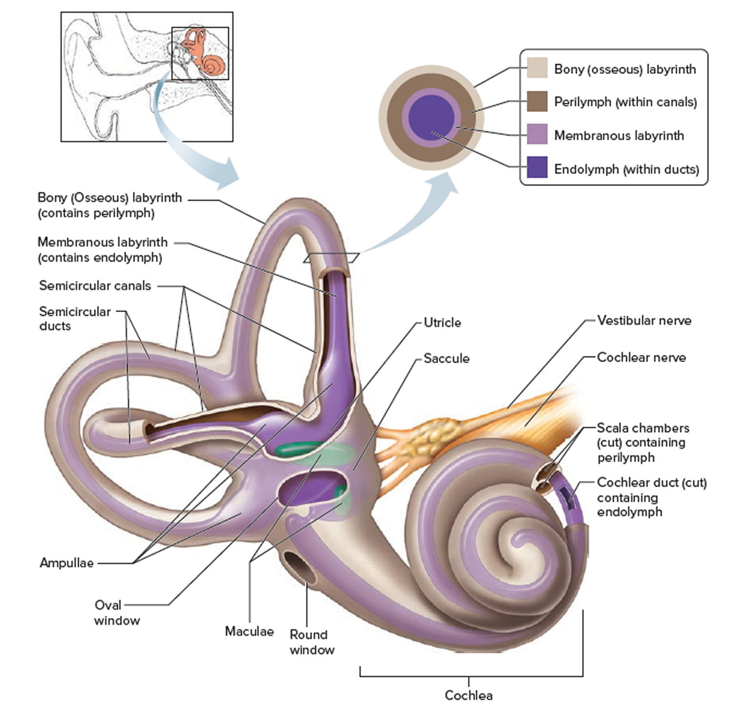
The ear also helps in balancing the body. The human ear allows us to feel the effect of gravity that is known as stationary balance and it also helps to feel the acceleration that is known as dynamic balance. The utricle and saccule provide a static balance. Dynamic balance is provided by semi-circular canals.
How The Ear Works Step by Step Brief Explanation
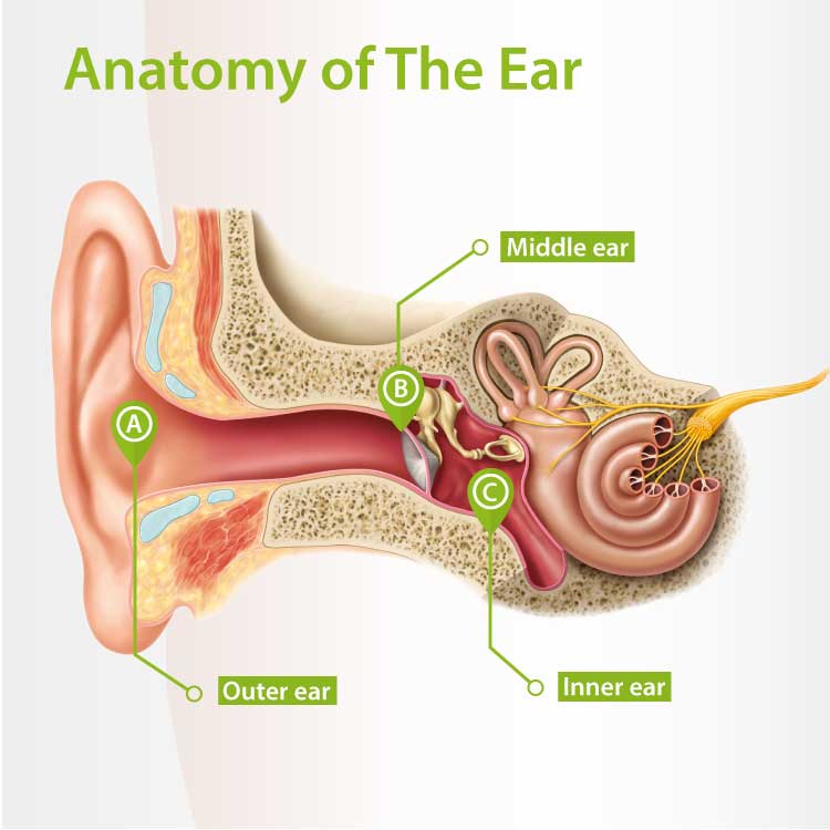
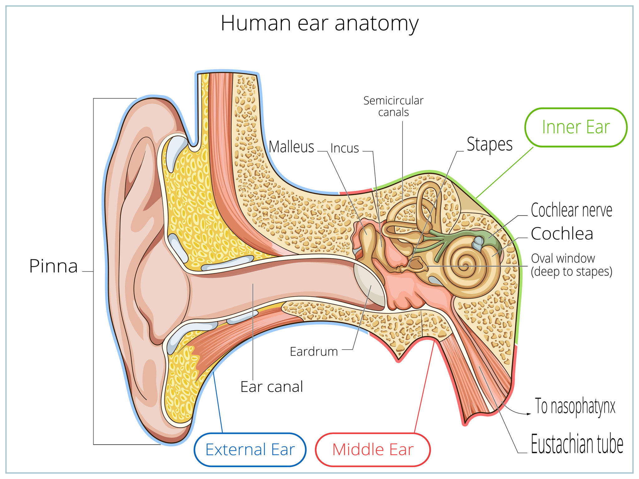
The ear is anatomically divided into three portions: External ear Middle ear Internal ear This mixture of bones, nerves, vessels, membranes, and muscles that make up the ear will be described in this article. Contents External ear Auricle External acoustic meatus Tympanic membrane Muscles of the external ear Vasculature of the external ear

1 Diagram showing the structure of the human ear, detailing the parts... Download Scientific
The Anatomy of the Ear Organs of human hearing are located on either side of the head By Mark Gurarie Updated on June 07, 2022 Medically reviewed by John Carew, MD Table of Contents Anatomy Function Associated Conditions Tests Essential for hearing and balance, each ear has an intricate structure of bones, nerves, and muscles.
How You Hear Northland Audiology
Helix: The outermost curvature of the ear, extending from where the ear joins the head at the top to where it meets the lobule. The helix begins the funneling of sound waves into the ear; Fossa, superior crus, inferior crus, and antihelix: These sections make up the middle ridges and depressions of the outer ear. The superior crus is the first ridge that emerges moving in from the helix.

Ear Diagram Helix Human Anatomy diagram
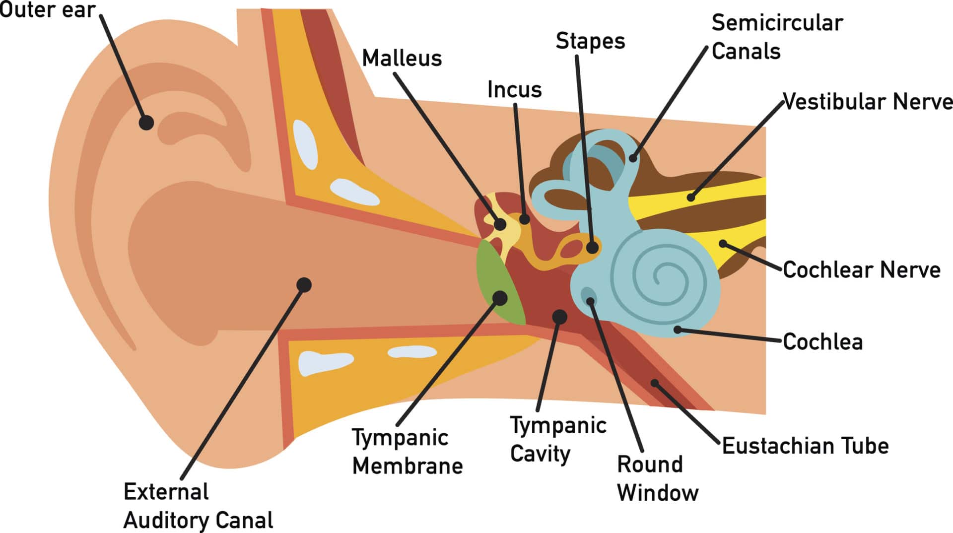
Structure The ear is made up of the outer ear, middle ear, and inner ear. The inner ear consists of the bony labyrinth and membranous labyrinth. The bony labyrinth comprises three components: Cochlea: The cochlea is made of a hollow bone shaped like a snail and divided into two chambers by a membrane.
Structure and Function of Human Ear with Diagram Teachoo
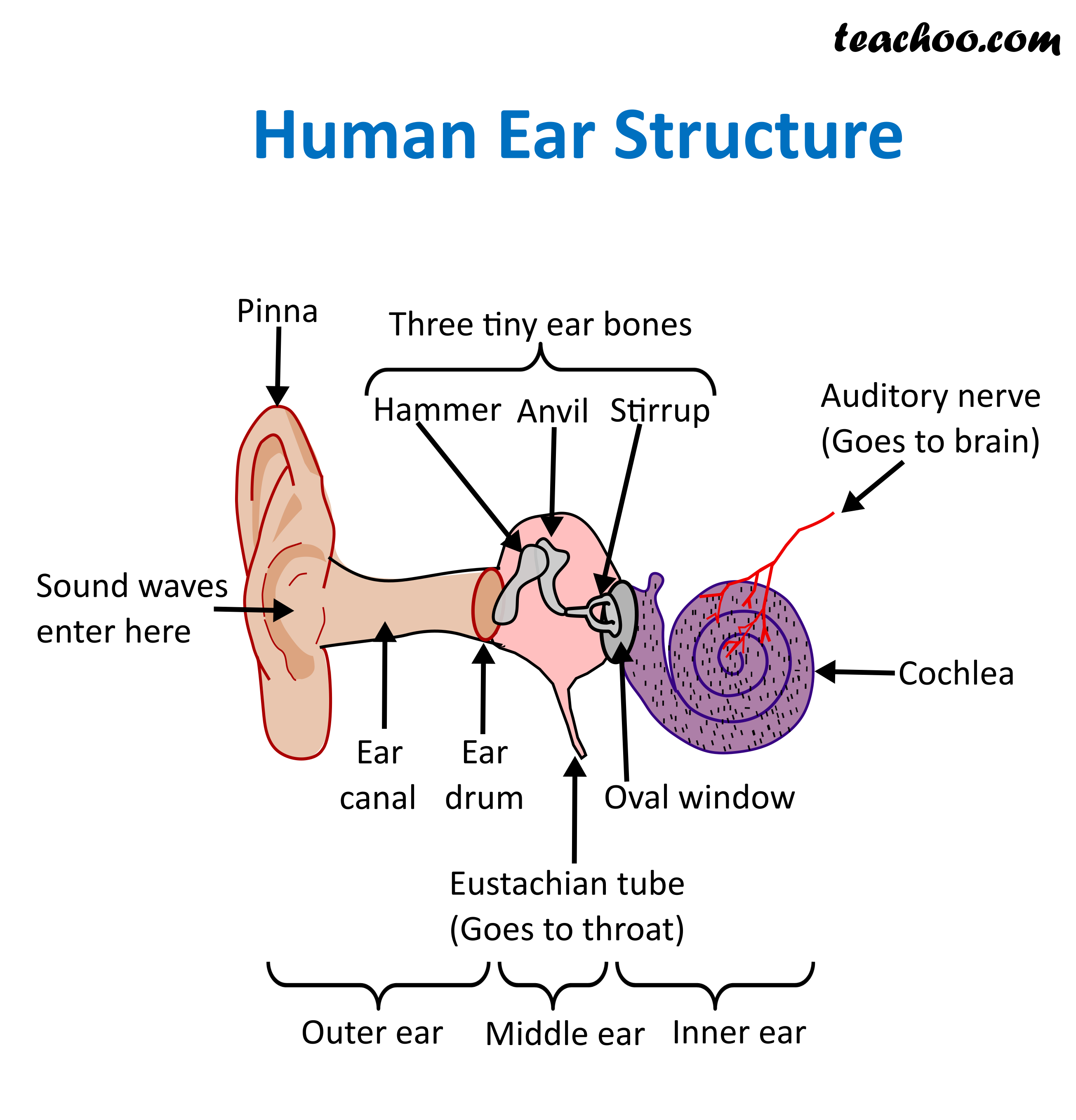
So as the air vibrates even the ear drum starts vibrating. Just like the skin of a drum. And as you can, the ear drum also separates the outer ear from the middle ear. This brings us to the middle ear. The middle ear consists of the three tiniest bones of the human body. And they're together the are called the ossicles. And they have pretty.

How The Ear Works
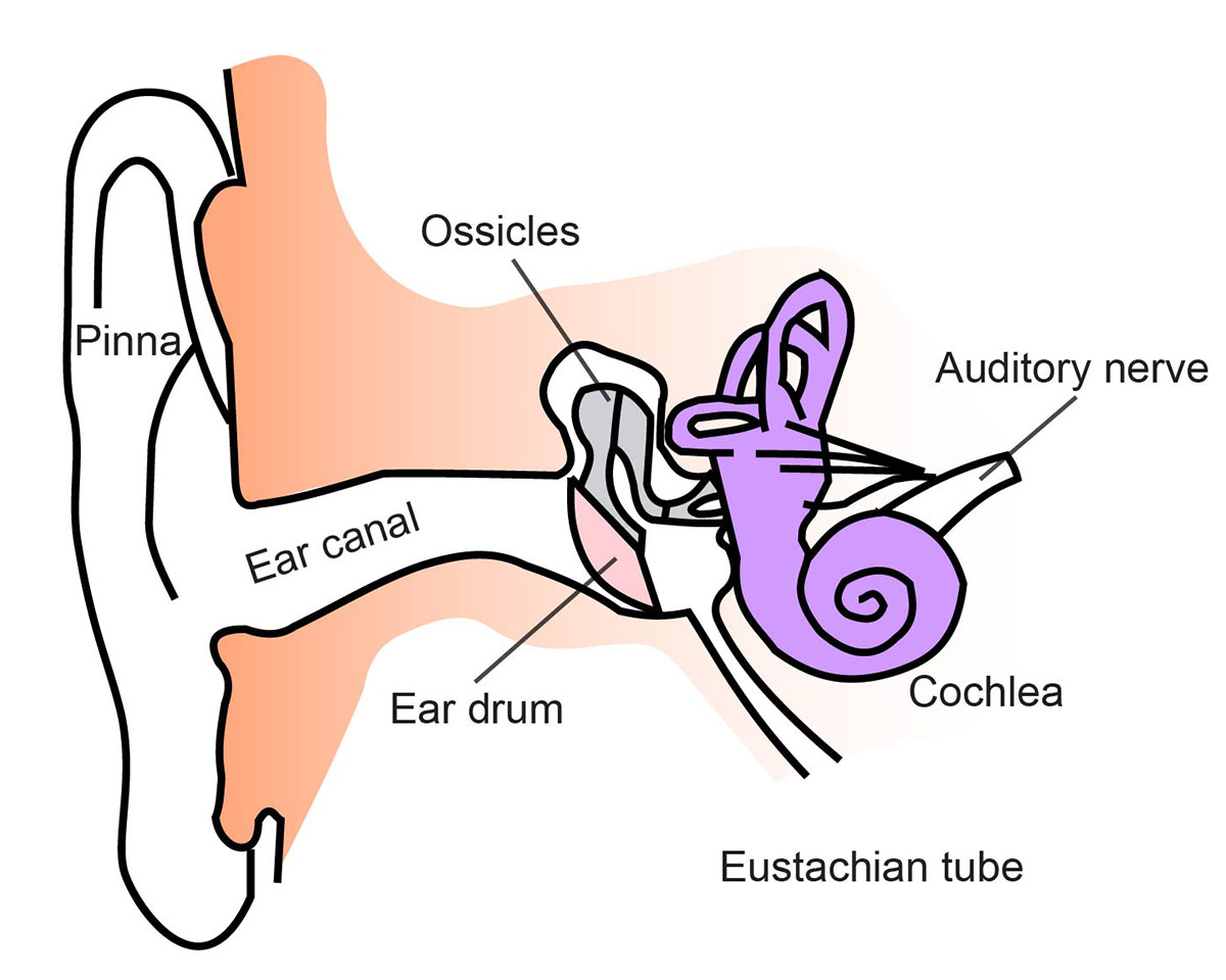
The human ear consists of three parts—the outer ear, middle ear and inner ear. [6] The ear canal of the outer ear is separated from the air-filled tympanic cavity of the middle ear by the eardrum.
Ear infections explained Dr Mark McGrath
The ear canal, or auditory canal, is a tube that runs from the outer ear to the eardrum. The ear has outer, middle, and inner portions. The ear canal and outer cartilage of the ear make.
Ear Anatomy Causes of Hearing Loss Hearing Aids Audiology
Diagram of Ear Human ear is a sense organ responsible for hearing and body balance. The outer ear receives the sound waves and transmits them down the ear canal to the eardrum. This causes the eardrum to vibrate and sound is produced. The diagram of the ear is important from Class 10 and 12 perspectives and is usually asked in the examinations.
Human Ear Anatomy Parts of Ear Structure, Diagram and Ear Problems
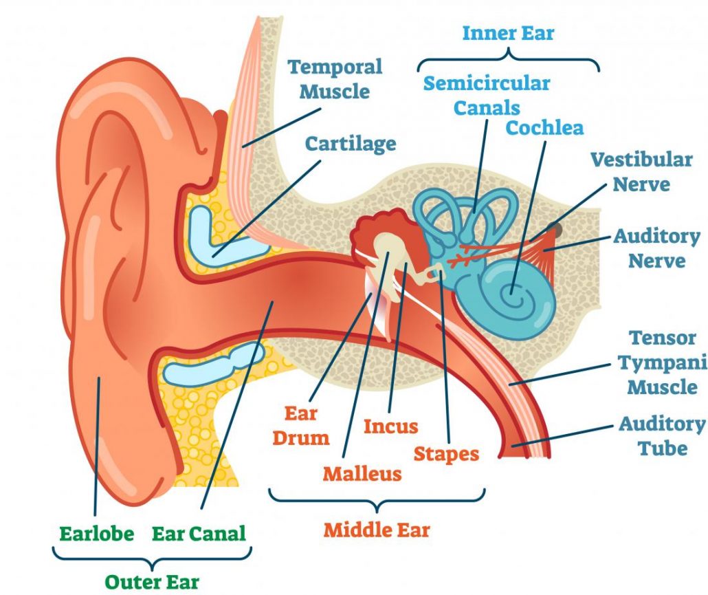
The human ear consists of three parts: External ear Middle ear Internal ear Human Ear Parts The human ear parts are explained below: External Ear The external ear is further divided into the following parts: Auricle (Pinna) The auricle comprises a thin plate of elastic cartilage covered by a layer of skin.
Ear Anatomy Causes of Hearing Loss Hearing Aids Audiology
The Human Ear www.TurnItToTheLeft.com. If the noise is too loud, walk away, turn it down (Turn it to the Left), or use ear plugs. pinna ear canal ear drum hammer anvil stirrup Eustachian tube (connects to the nose) cochlea semicircular canals nerves (connect to the brain) Directions: Color in the diagram below using a different color for.
